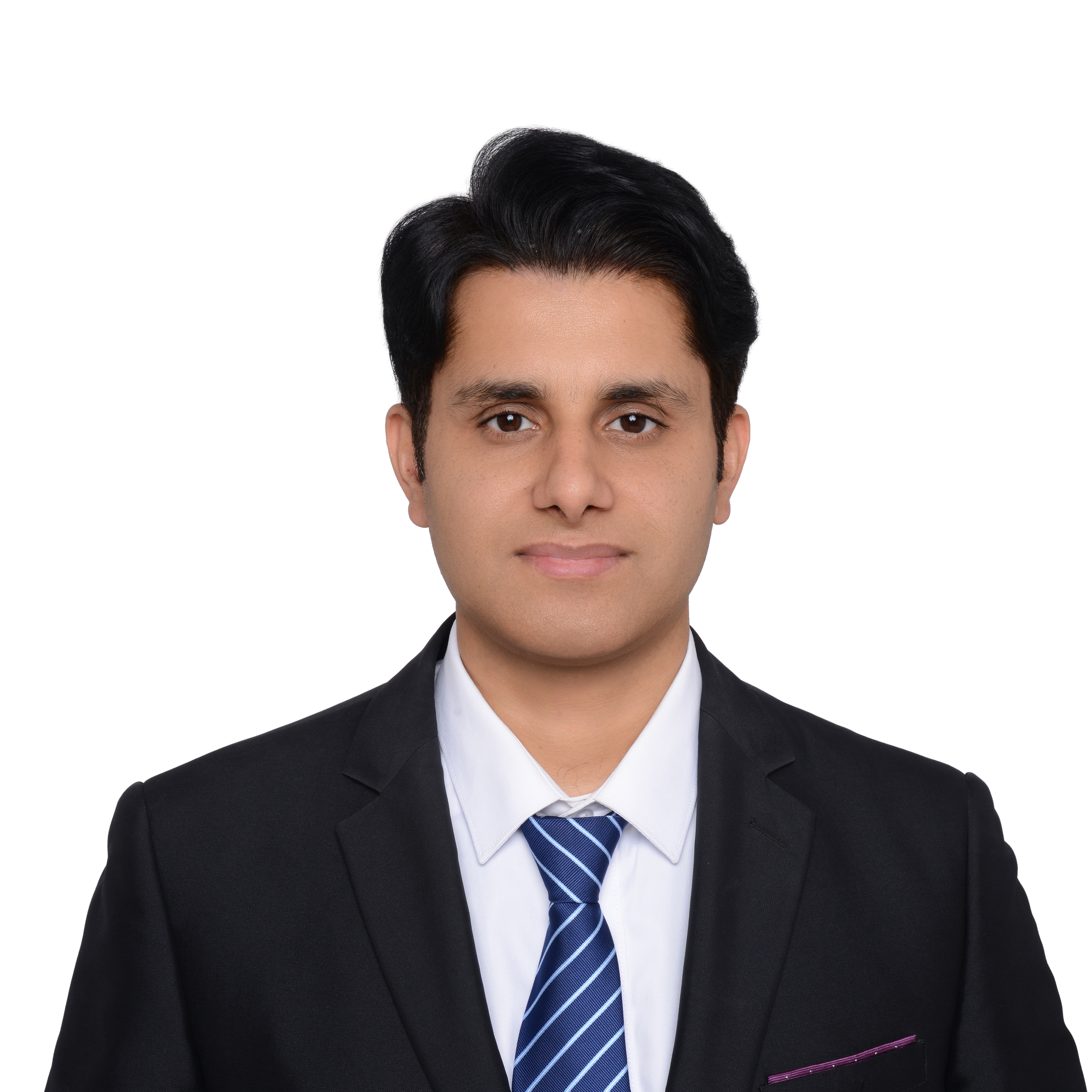
Dr. Muhammad Aamir
Session Speaker
Dr. Muhammad Aamir is an Associate Professor and Chutian Scholar at Huanggang Normal University, China, where he leads research in artificial intelligence and medical imaging. He holds a Ph.D. in Computer Science and Technology from Sichuan University (2019) and an M.Eng. in Software Engineering from Chongqing University (2014). Recognized among the Top 2% of Scientists Worldwide (2025) by Stanford University, Dr. Aamir specializes in deep learning for medical image analysis, particularly brain tumor detection and cancer diagnosis using MRI and CT imaging. He has published extensively in high-impact journals including Scientific Reports, IEEE Access, and Biomedical Signal Processing and Control, with over 80 publications to his credit. Dr. Aamir has secured multiple research grants as Principal Investigator and serves on the editorial boards of several international journals. He has edited four books on deep learning and medical signal processing, delivered numerous keynote speeches at international conferences, and organized workshops on AI applications in healthcare. Beyond research, he has held teaching positions in China and Pakistan, worked as a software support engineer in the industry, and actively contributes to academic communities through peer review and conference organization.
Title: AI in Healthcare: Deep Learning for Brain Tumor Classification from MRI Abstract: Brain tumors can be fatal if not detected early, and manual diagnosis depends on radiological expertise that is not uniformly available. This keynote situates automated brain tumor classification within the broader adoption of deep learning across healthcare, and presents a complete pipeline for classifying gliomas, meningiomas and pituitary tumors from MRI. The method proceeds in four stages. MRI scans are first enhanced to address the low contrast, non-uniform illumination and artefacts typical of multi-modality acquisition. Two pre-trained deep networks then extract complementary feature representations, which are combined into a single hybrid feature vector using partial least squares fusion. Candidate tumor locations are generated through agglomerative clustering and ranked by an edge-based scoring function, and the resulting proposals are aligned to a fixed size before classification. Evaluated on a public benchmark of 3,064 contrast-enhanced MRI scans from 233 patients, the approach achieves 98.95% overall accuracy, with per-class accuracies of 98.30%, 98.72% and 99.37%, and a detection recall of 95.4% using only 100 proposals per image. The talk concludes by discussing the practical requirements for translating classification performance of this kind into clinical use, including validation protocol, external evaluation, and integration with radiological workflow.